



Early skin cancer diagnosis: Changing moles and wounds need attention
Early assessment of growing, bleeding or non-healing skin lesions can support more limited treatment and help preserve patients’ quality of life.


Bilge Türk | MediCenter TV
ANKARA, TÜRKİYE — Skin lesions that grow, bleed, change appearance or fail to heal within several weeks should be examined promptly, as early diagnosis may allow skin cancer to be treated with more limited procedures.
Skin cancer is among the commonly encountered types of cancer worldwide, but its early signs can sometimes be mistaken for an ordinary mark, mole or persistent wound. Dr. Seval Karasatı, who works in dermatology, says lesions that remain for a long time or change over time should be assessed by a dermatologist.
Skin cancer develops when genetic changes in skin cells lead to uncontrolled cell growth. Prolonged or intense exposure to ultraviolet radiation is an important risk factor for many forms of the disease. Genetic characteristics, immune system status and certain environmental factors may also contribute to its development.
Because changes can appear on the skin’s surface, people may have an opportunity to seek medical advice at an early stage. However, not every skin cancer begins with an obvious or easily recognised symptom. Some lesions may initially be small, flat, painless or appear harmless.


Different skin cancers may produce different warning signs
Skin cancers can originate from different types of cells. The most frequently encountered forms include basal cell carcinoma, squamous cell carcinoma and melanoma. Their growth patterns, potential to spread and treatment approaches differ.
Basal cell carcinoma is one of the most common forms of skin cancer. It can develop on sun-exposed areas such as the face, ears, neck and scalp. It may appear as a shiny or pearl-like bump, a pink patch, a superficial wound or a lesion that seems to heal before opening again.
This form of cancer generally grows slowly, and spread to distant organs is rare. If it remains untreated, however, it can extend into surrounding tissue. Lesions located on sensitive areas such as the face may result in substantial tissue loss when treatment is delayed.
Squamous cell carcinoma can appear as a firm, crusted or scaly area or as a wound that does not heal. It may occur on parts of the head, neck and trunk exposed to intense sunlight. The lower lip may also be affected more frequently among people who use tobacco.


Some squamous cell carcinomas can spread into surrounding tissues or lymph nodes. Persistent crusting, growth or failure to heal therefore requires timely medical assessment.
Melanoma develops from pigment-producing cells known as melanocytes. Compared with some other skin cancers, it may have a greater potential to spread. A change in an existing mole or the appearance of a new pigmented lesion may be among its initial signs.
Melanoma is not always black or dark brown. Some lesions may appear pink, red or skin-coloured. For this reason, monitoring only dark-coloured moles is not sufficient.
Early diagnosis can influence the extent of treatment
One of the principal advantages of early diagnosis is the possibility of identifying skin cancer while it remains confined to a relatively small area. In melanoma, the depth of the tumour and whether it has spread are important factors in determining treatment and the course of the disease.
Many skin cancers detected at an early stage can be treated successfully with surgery. When the disease progresses, patients may require wider surgical procedures, different treatment methods and more extensive follow-up.
A lesion should not be considered harmless simply because it causes no pain. Skin cancers may be painless in their early stages, meaning that pain alone is not a reliable measure of whether a skin change requires attention.
Wounds that do not heal within several weeks, areas that repeatedly develop crusts, lesions that bleed without an obvious cause and rapidly growing skin formations may require examination by a dermatologist. A noticeable change in an existing mole can also justify professional assessment.
Early diagnosis may additionally reduce functional and cosmetic problems resulting from treatment. Lesions affecting the nose, eyelids, lips or ears may be treated with less tissue loss when they are identified and addressed at an earlier stage.
Some people may face a higher risk
Skin cancer can occur in people of different ages and skin tones. Nevertheless, certain characteristics and forms of exposure may increase the likelihood of developing the disease.
People with fair skin, light-coloured eyes, numerous moles or a tendency to burn easily in sunlight may have a higher risk of particular skin cancers. Severe sunburn during childhood may also affect the risk of melanoma later in life.
Cumulative ultraviolet exposure is an important concern for people who have worked outdoors for many years. Individuals employed in agriculture, construction and similar occupations should pay particular attention to sun-protection habits.
The use of tanning beds may increase the risk of melanoma and certain other skin cancers. The belief that artificial ultraviolet radiation provides a safe method of tanning is not scientifically accurate.
A family history of melanoma and a previous diagnosis of skin cancer are also relevant when individual risk is assessed. Some skin cancers may occur more frequently in people taking immune-suppressing medicines following an organ transplant.
People with darker skin can also develop skin cancer. Certain melanomas may appear on the palms of the hands, the soles of the feet or beneath the nails. Skin colour alone therefore does not mean that a person has no risk of the disease.
Dermatological examination supports an accurate diagnosis
When skin cancer is suspected, a dermatologist assesses the lesion’s size, colour, borders and surface characteristics. Information about how long it has been present and whether it has changed over time is also considered.
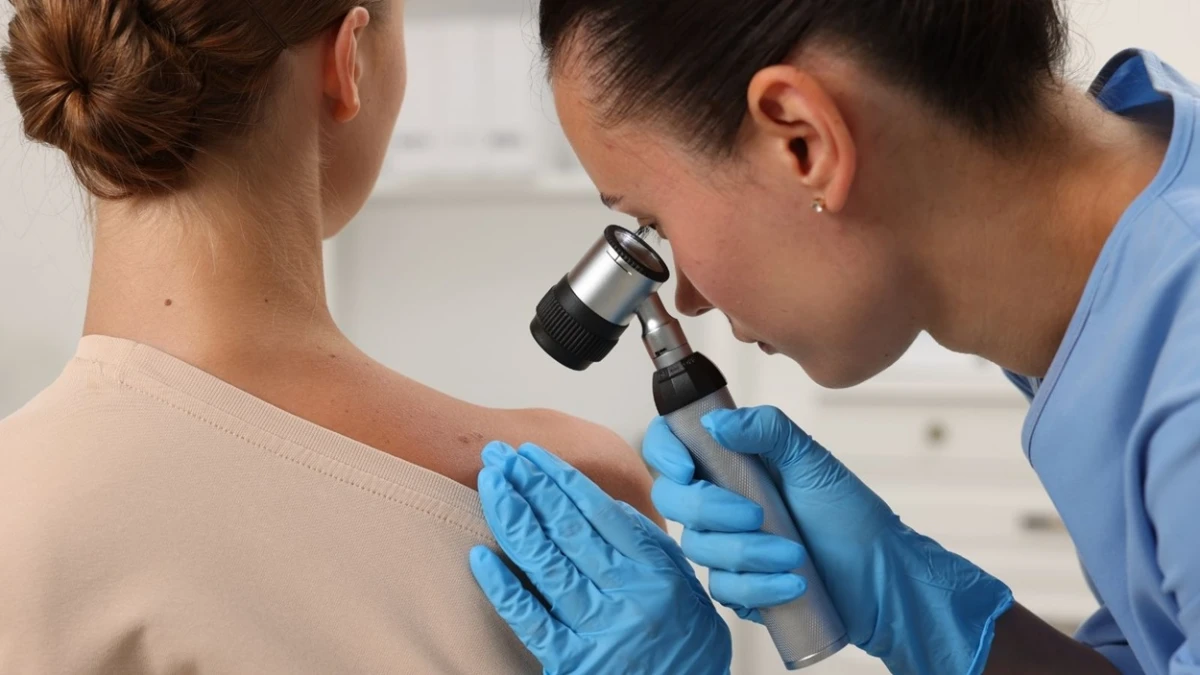
Dermoscopy is an additional method used to examine some pigmented and non-pigmented lesions. It enables the dermatologist to inspect structures within the skin that may not be easily visible to the naked eye.
Although dermoscopic examination can help identify suspicious lesions, it does not provide a definitive diagnosis in every case. When necessary, the dermatologist may recommend taking a biopsy or surgically removing the lesion.
Pathological examination is one of the principal methods used to confirm a skin cancer diagnosis. The findings provide information about the type and characteristics of the cancer and, when required, help determine the disease stage.
Treatment is planned according to the tumour’s type, size, location and depth, along with the patient’s general health. Possible approaches include surgical excision, Mohs micrographic surgery, local treatments for certain superficial lesions and systemic treatment for advanced disease.
Treatment should not be regarded as a procedure intended only to improve the appearance of the skin. Its primary purpose is to treat cancerous tissue appropriately and prevent the disease from progressing.
Sun protection and regular skin awareness should work together
Reducing unnecessary ultraviolet exposure is an important part of skin cancer prevention. Avoiding prolonged unprotected exposure during periods of intense sunlight is among the main protective measures.
Broad-spectrum sunscreen with a suitable protection factor should form part of a wider sun-protection routine. Hats, sunglasses and protective clothing can provide additional physical protection.
Using sunscreen does not mean that a person can remain in the sun for an unlimited period. The product should be applied in the recommended amount and reapplied when necessary.
Knowing the usual appearance of one’s skin may make it easier to notice new lesions or changes in existing moles. Monitoring wounds that do not heal, growths that become larger and areas that bleed without an apparent reason can encourage earlier medical assessment.
Self-examination does not replace a professional skin examination. For people in higher-risk groups, the appropriate frequency of dermatological checks should be determined individually.
Dr. Karasatı says early detection is important not only for treatment success but also for preserving quality of life. Not every changing mole or skin mark is cancerous, but lesions that grow, bleed, change appearance or fail to heal should be examined by a qualified specialist.
Skin cancer awareness should help people understand warning signs without creating unnecessary anxiety. Sun protection, awareness of personal risk factors and the timely examination of suspicious skin changes remain central to protecting long-term skin health.
MediCenterTV.com
 Like
0
 Dislike
0
 Love
0
 Funny
0
 Wow
0
 Sad
0
 Angry
0

Bilge Türk, MediCenterTV.com editörü, gazeteci ve yazardır. Sağlık, yaşam, bilim ve güncel gelişmeler üzerine içerikler hazırlayan Türk; doğru, anlaşılır ve toplum yararını gözeten habercilik anlayışıyla sağlık alanındaki gelişmeleri okuyuculara aktarmayı amaçlamaktadır.


















Comments (0)